
fluoroscopy guided injection
- Published
- in Guide
Fluoroscopy-guided injections involve using real-time X-ray imaging to precisely administer medications, enhancing accuracy and effectiveness in both diagnostic and therapeutic medical procedures.
1.1 Definition and Overview
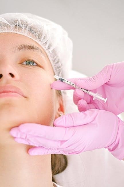
Fluoroscopy-guided injections are minimally invasive procedures that use real-time X-ray imaging to accurately deliver medications, such as steroids or anesthetics, to specific targets within the body. This technique enhances precision, ensuring the needle is placed correctly for diagnostic or therapeutic purposes. Widely used in pain management and musculoskeletal interventions, fluoroscopy provides immediate feedback, improving the efficacy of treatments and minimizing complications. It is a valuable tool in modern medicine, balancing accuracy with patient safety.
1.2 Historical Background
Fluoroscopy-guided injections trace their origins to the discovery of X-rays by Wilhelm Conrad Röntgen in 1895. Initially used for diagnostic imaging, fluoroscopy evolved to guide medical interventions. The mid-20th century saw its application in pain management and orthopedics. Technological advancements in the 1980s and 1990s improved image quality and safety, making fluoroscopy a standard tool for precise needle placement. This historical progression highlights its transformation from a diagnostic aid to a critical component in minimally invasive therapies, enhancing treatment accuracy and patient outcomes.
1.3 Importance in Medical Practice
Fluoroscopy-guided injections are crucial in modern medicine, offering precise needle placement during procedures. They enhance diagnostic accuracy, allowing clinicians to identify pain sources and deliver targeted therapies. This technique minimizes complications, improves treatment outcomes, and reduces recovery times. Its real-time imaging capability ensures accurate medication delivery, making it invaluable in pain management, orthopedics, and interventional radiology. The integration of fluoroscopy in medical practice has revolutionized minimally invasive treatments, providing both therapeutic and diagnostic benefits with high efficiency.

Fluoroscopy: The Core Technology
Fluoroscopy uses continuous X-ray beams to create real-time images, enabling precise guidance during injections. This technology is essential for visualizing dynamic body structures, ensuring accurate needle placement and minimizing risks.
2.1 Principles of Fluoroscopy
Fluoroscopy operates by transmitting continuous X-ray beams through a patient’s body, capturing real-time images. These images are enhanced using image intensifiers and cameras, allowing for dynamic visualization. The technology enables precise tracking of needle movements, ensuring accurate placement during injections. Its ability to provide immediate feedback makes it invaluable for guiding medical procedures, reducing complications, and improving outcomes in various clinical settings.
- Real-time imaging enhances procedural accuracy.
- Continuous X-ray beams capture dynamic body structures.
- Image intensifiers improve visibility for precise needle placement.
2.2 Equipment and Setup
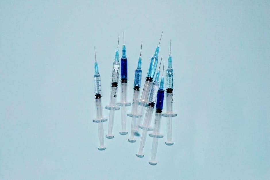
Fluoroscopy-guided injections require specialized equipment, including a fluoroscope, image intensifier, and high-resolution monitor. The fluoroscope emits a continuous X-ray beam, while the intensifier enhances image quality. A radiolucent patient table allows unobstructed X-ray transmission. Additional components include a control panel for adjusting settings and a camera to capture images. Proper shielding and protective gear are essential to minimize radiation exposure for both patients and operators, ensuring safety during procedures.
- Fluoroscope with image intensifier and camera.
- High-resolution display monitor.
- Radiolucent patient table.
- Lead shielding for radiation protection.
2.3 Radiation Safety Considerations
Radiation safety is critical in fluoroscopy-guided injections to minimize exposure risks. Operators and patients must wear lead aprons, thyroid shields, and use dosimeters to monitor exposure. The ALARA principle (As Low As Reasonably Achievable) guides dose reduction. Shielding and collimation techniques are employed to limit unnecessary radiation. Regular equipment calibration and proper training for operators further enhance safety protocols, ensuring procedures are conducted with minimal risk to all involved while maintaining diagnostic accuracy.
- Use of lead aprons and thyroid shields.
- Dosimeters for exposure monitoring.
- Collimation to reduce scatter radiation.
- Regular equipment maintenance.

Types of Fluoroscopy-Guided Injections
Fluoroscopy-guided injections include epidural steroid injections, facet joint injections, and joint or soft tissue injections, each targeting specific anatomical areas for pain relief and diagnostic purposes.
- Epidural steroid injections for spinal pain.
- Facet joint injections for spinal facet pain.
- Joint and soft tissue injections for inflammation.
3.1 Epidural Steroid Injections
Epidural steroid injections are a common fluoroscopy-guided procedure to treat spinal pain and inflammation. They involve injecting a mixture of steroids and anesthetics into the epidural space surrounding the spinal cord. This method is often used to alleviate pain caused by conditions such as herniated discs, spinal stenosis, or nerve root compression. The use of fluoroscopy ensures precise needle placement, minimizing risks and improving effectiveness. These injections provide targeted relief, reducing inflammation and improving mobility for patients with chronic pain.
By delivering medication directly to the affected area, epidural injections offer a localized treatment approach, reducing systemic side effects. This procedure is particularly beneficial for patients who have not responded to conservative treatments like physical therapy or oral medications. Fluoroscopy guidance enhances accuracy, ensuring the medication reaches the intended location for optimal results.
3.2 Facet Joint Injections
Facet joint injections are fluoroscopy-guided procedures used to diagnose and treat pain originating from the facet joints, small stabilizing structures in the spine. These injections typically involve administering a combination of steroids and anesthetics into the joint or surrounding nerves to reduce inflammation and alleviate pain. Commonly used for conditions like facet syndrome or spinal pain, this method ensures precise needle placement under fluoroscopic imaging, enhancing therapeutic outcomes and minimizing complications.
The procedure is particularly effective for patients experiencing chronic back pain, as it targets the specific source of discomfort. Fluoroscopy allows real-time visualization, ensuring accurate delivery of medication directly to the affected facet joint. This targeted approach can significantly improve mobility and reduce pain in individuals with spinal disorders, making it a valuable option in pain management strategies.
3.3 Joint and Soft Tissue Injections
Fluoroscopy-guided joint and soft tissue injections are procedures used to treat pain and inflammation in joints, tendons, and ligaments. These injections often involve corticosteroids or anesthetics delivered precisely to the affected area under real-time imaging. Common applications include joint aspirations to remove fluid for diagnostic purposes and therapeutic injections to reduce inflammation in conditions like arthritis or tendinitis. Fluoroscopy ensures accurate needle placement, enhancing the effectiveness of the treatment while minimizing risks to surrounding tissues.
This technique is particularly useful for targeting deep or hard-to-reach areas, such as hip or shoulder joints, where blind injections might be less accurate. By providing clear visualization, fluoroscopy helps minimize complications and improves patient outcomes, making it a reliable choice for both diagnostic and therapeutic interventions in musculoskeletal care.

Indications for Fluoroscopy-Guided Injections
Fluoroscopy-guided injections are primarily used for diagnostic purposes, therapeutic interventions, and pain management, targeting specific areas to deliver precise medication and reduce inflammation or discomfort effectively.
4.1 Diagnostic Purposes
Fluoroscopy-guided injections are utilized for diagnostic purposes to identify the source of pain or inflammation. By injecting contrast agents or medications under real-time imaging, healthcare providers can pinpoint specific areas causing discomfort. This method is particularly effective in assessing joint or soft tissue abnormalities, allowing for precise diagnosis and tailored treatment plans. It ensures accurate delivery of diagnostic agents, enhancing the ability to visualize and confirm conditions such as joint inflammation or nerve-related pain.
4.2 Therapeutic Applications
Fluoroscopy-guided injections are widely used for therapeutic purposes, delivering medications directly to targeted areas. This method ensures precise administration of corticosteroids, anesthetics, or other therapies to reduce inflammation, relieve pain, and improve mobility. Common applications include treating arthritis, tendinitis, and degenerative joint conditions. Real-time imaging allows for accurate needle placement, enhancing the effectiveness of treatments like joint injections or nerve blocks, while minimizing risks to surrounding tissues.
4.3 Pain Management
Fluoroscopy-guided injections are invaluable in pain management, particularly for chronic conditions like arthritis, herniated discs, and spinal stenosis. By enabling precise delivery of anesthetics or steroids, these injections target pain sources effectively, reducing inflammation and providing relief. Common procedures include epidural steroid injections for spinal pain and facet joint injections for localized discomfort. This method ensures accurate needle placement, minimizing complications and enhancing therapeutic outcomes, making it a preferred approach for managing persistent pain in various musculoskeletal and neural conditions.

Contraindications and Risks
Contraindications include allergies to contrast agents, active infections, or unstable medical conditions. Risks involve radiation exposure, bleeding, or nerve damage, requiring careful patient screening.
5.1 Absolute Contraindications
Absolute contraindications for fluoroscopy-guided injections include severe allergies to contrast agents, active infections, or pregnancy. Conditions like unstable medical states or inability to cooperate during the procedure also apply. These factors pose significant risks, making the procedure unsafe under any circumstances. Proper patient screening is essential to identify and exclude such cases, ensuring the procedure’s safety and effectiveness.
5.2 Relative Contraindications
Relative contraindications for fluoroscopy-guided injections include conditions that may increase procedure risks but are not absolute barriers. These include bleeding disorders, unstable medical conditions, or severe allergies to medications. Additionally, patient inability to remain still or follow instructions during the procedure can complicate outcomes. Other factors, such as pregnancy or use of certain medications like blood thinners, may also require cautious consideration. These cases necessitate careful evaluation to weigh potential benefits against risks.
5.3 Potential Complications
Potential complications of fluoroscopy-guided injections include bleeding, infection, or nerve damage at the injection site. Radiation exposure poses risks, especially with prolonged procedures. Allergic reactions to medications or contrast agents are rare but possible. In rare cases, accidental injection into blood vessels or surrounding tissues can occur, potentially causing systemic effects. These risks are minimized with precise technique, sterile conditions, and proper patient screening. Discussing these complications with a healthcare provider is essential for informed decision-making.

Patient Preparation and Screening
Patient preparation involves thorough screening, education, and post-care instructions to ensure safety and effectiveness during fluoroscopy-guided injections.
6.1 Pre-Procedure Screening
Pre-procedure screening for fluoroscopy-guided injections involves assessing the patient’s medical history, current medications, and allergies. It ensures the procedure’s safety and suitability. Key steps include reviewing imaging studies, evaluating kidney function, and confirming the absence of contraindications. Patients are also informed about potential risks and benefits, and written consent is obtained. Additionally, radiation exposure risks are discussed, especially for pregnant or sensitive individuals. This step ensures personalized care and minimizes complications.
6.2 Patient Education
Patient education is crucial for ensuring understanding and cooperation during fluoroscopy-guided injections. Patients should be informed about the procedure’s purpose, benefits, and potential risks. Clear instructions on preparation, such as fasting or medication adjustments, are provided. The use of fluoroscopy for real-time guidance is explained to reassure patients. Discussing expected outcomes and recovery instructions helps manage expectations. Effective communication ensures patients are comfortable and informed, promoting a positive experience and adherence to post-procedure care.
6.4 Post-Procedure Care
Post-procedure care for fluoroscopy-guided injections focuses on monitoring and recovery. Patients are observed for potential complications like bleeding or swelling. They are advised to rest the treated area and avoid strenuous activities for a specified period. Pain management options may be provided, and follow-up appointments are scheduled to assess efficacy. Clear instructions on wound care and signs of complications are emphasized to ensure patient safety and optimal recovery outcomes.
Benefits of Fluoroscopy Guidance
Fluoroscopy guidance offers enhanced accuracy, reduced complications, and cost-effectiveness, making it a valuable tool in medical procedures.
7.1 Accuracy and Precision
Fluoroscopy guidance significantly enhances the accuracy and precision of injections by providing real-time imaging, ensuring the needle is placed exactly where needed. This reduces the risk of hitting vital structures and improves the effectiveness of treatments. The ability to visualize the injection site during the procedure minimizes blind injections and enhances therapeutic outcomes, making fluoroscopy a reliable tool for precise medical interventions.
7.2 Reduced Complication Rates
Fluoroscopy-guided injections significantly reduce complication rates by enabling precise needle placement under real-time visualization. This minimizes the risk of inadvertently damaging nearby structures, such as nerves or blood vessels. The ability to monitor the injection process also reduces the likelihood of infection or other procedural complications. These advantages make fluoroscopy-guided injections safer and more reliable compared to blind or unguided techniques, particularly for patients with complex anatomy or medical conditions that increase procedural risks.
7.3 Cost-Effectiveness
Fluoroscopy-guided injections are cost-effective due to their efficiency and precision. The use of real-time imaging reduces the need for repeat procedures and additional imaging, lowering overall healthcare costs. Fluoroscopy is widely available, low-cost, and quick, making it a practical choice for many medical practices. By minimizing complications and improving accuracy, these injections contribute to reduced healthcare expenditure while providing effective patient care, making them a valuable option for both providers and patients.

Limitations of Fluoroscopy-Guided Injections
Fluoroscopy-guided injections have limitations, including radiation exposure concerns, limited visualization of soft tissues, and accessibility issues in certain clinical settings, affecting their universal applicability.
8.1 Radiation Exposure Concerns
Fluoroscopy-guided injections involve radiation exposure, posing potential risks to patients and providers. Prolonged procedures increase cumulative dose, raising concerns about cancer and genetic risks. Modern systems use low-dose settings, but repeated exposure remains a concern. Protective gear and monitoring are essential to minimize risks. The benefits often outweigh the risks, but careful patient selection and dose monitoring are critical to ensure safety. Radiation safety protocols are vital to mitigate long-term health impacts.
8.2 Availability and Accessibility
Fluoroscopy-guided injections are widely available in healthcare settings, particularly in hospitals and specialized clinics. The procedure’s cost-effectiveness and quick execution make it accessible for many patients. However, availability can vary depending on geographic location, with urban areas typically having better access than rural regions. Additionally, the need for specialized equipment and trained professionals may limit accessibility in some underserved areas, highlighting disparities in healthcare resources and expertise.
8.3 Technical Challenges
Fluoroscopy-guided injections require specialized equipment and skilled operators, posing technical challenges. Equipment limitations, such as image resolution and radiation exposure constraints, can affect procedure accuracy. Additionally, maintaining precise needle placement under real-time imaging demands high operator expertise. Technical difficulties may also arise from patient movement or anatomical complexity, potentially complicating the injection process. Ensuring optimal image quality while minimizing radiation exposure remains a critical technical challenge in these procedures.

Comparison with Other Imaging Techniques
Fluoroscopy-guided injections are compared to ultrasound, CT, and MRI for accuracy and efficiency. Fluoroscopy offers real-time imaging, making it ideal for dynamic procedures, though it may lack in soft tissue detail compared to other modalities.
9.1 Ultrasound-Guided Injections
Ultrasound-guided injections are a popular alternative to fluoroscopy, offering real-time imaging without radiation exposure. This method provides excellent visualization of soft tissues, making it ideal for superficial injections. Ultrasound is portable, cost-effective, and does not expose patients or operators to ionizing radiation, enhancing safety. However, it is highly operator-dependent and may lack the depth penetration required for deeper structures. Despite these limitations, ultrasound is widely used for its precision and non-invasive nature, making it a valuable tool in modern medicine.
9.2 CT-Guided Injections
CT-guided injections use computed tomography to provide high-resolution images, allowing precise needle placement in complex anatomical areas. This method is advantageous for deep tissue injections due to its superior soft tissue differentiation. However, it involves higher radiation exposure and greater costs compared to fluoroscopy. CT guidance is particularly useful in cases where fluoroscopy or ultrasound are insufficient, offering enhanced accuracy for challenging procedures. Despite its benefits, the higher radiation dose and limited availability make it less common for routine injections.
9.3 MRI-Guided Injections
MRI-guided injections offer exceptional soft tissue visualization without radiation, utilizing magnetic fields to guide needles precisely. This method is ideal for injections in sensitive or complex anatomical regions. While it provides superior image quality compared to fluoroscopy or CT, MRI’s limited availability and higher costs restrict its use. Additionally, longer procedure times and compatibility issues with certain medical implants can hinder its application, making it less common for routine injections despite its diagnostic advantages.
Future Trends in Fluoroscopy Technology
Advancements in fluoroscopy include improved image resolution, AI integration for real-time analytics, and enhanced radiation safety features, making procedures more precise, efficient, and patient-friendly.
10.1 Advancements in Image Quality
Fluoroscopy-guided injections are benefiting from advancements in image quality, including higher resolution and better contrast. Improved detector sensitivity enhances visualization of soft tissues and small structures, reducing the need for retakes. These enhancements improve diagnostic accuracy and precision during injections, ensuring better patient outcomes and minimizing complications. Additionally, better image clarity aids in identifying intricate anatomical details, making procedures safer and more effective for both patients and practitioners.
10.2 Integration with AI and Robotics
The integration of AI and robotics in fluoroscopy-guided injections is revolutionizing the field by enhancing precision and efficiency. AI algorithms can analyze images in real-time, improving accuracy and reducing radiation exposure. Robotic systems assist in needle placement, minimizing human error and improving consistency. These technologies also enable automated tracking of injected substances and provide real-time feedback, ensuring optimal outcomes. The combination of AI and robotics is expected to further elevate the safety and effectiveness of fluoroscopy-guided procedures, benefiting both patients and practitioners.
10.3 Enhanced Radiation Safety Features
Future advancements in fluoroscopy-guided injections emphasize enhanced radiation safety features. These include advanced dose-reduction technologies, real-time radiation monitoring, and automated exposure controls. Modern systems now incorporate pulsed X-ray beams and “last-image hold” features, minimizing radiation exposure without compromising image quality. Additionally, AI-driven algorithms optimize radiation dosage, ensuring patient and operator safety while maintaining procedural efficacy. These innovations align with growing demands for safer, more efficient medical imaging practices.
Fluoroscopy-guided injections represent a cornerstone in modern medical practice, offering precise and effective solutions for pain management and diagnostic procedures. By leveraging real-time imaging, these injections enhance accuracy, reduce complications, and improve patient outcomes. Despite the balance of benefits and risks, advancements in radiation safety and technology continue to expand their applications. As medicine evolves, fluoroscopy-guided injections remain a vital tool, bridging the gap between imaging and intervention for optimal care.